PET
PET stands for positron emission tomography and is a method for medical imaging diagnostics. The technique involves using radioactive drugs that emit a signal readable by a PET scanner. PET diagnostics can be used applied across various medical fields.
What is PET
PET is a tool that helps investigate patients with various diseases. These can include cancer, neurological diseases, cardiovascular diseases, or inflammatory conditions. The examination is conducted by injecting “target-seeking” molecules that are radioactively labelled, allowing tracking throughout the body. These molecules accumulate more in diseased tissue than in healthy tissue. The radiation emitted by these molecules is captured by a detector and converted into an image.
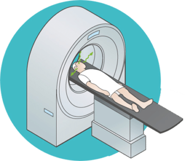
The actual examination takes place in a scanner, typically after a waiting period of up to three quarters of an hour for the molecules to distribute in the body. PET leverages the fact that the radioactive isotopes injected into the body are unstable. When they convert to stable isotopes, they emit particles known as positrons. These interact with electrons in the tissue and convert into gamma radiation. This radiation is emitted in two directions simultaneously. The scanner, which the patient lies in, has a ring of detectors that capture the gamma radiation, allowing precise localization of the source of the radiation in the body. By overlaying this information on a CT or MRI image, a picture of the diseased tissue can be created.
What is PET used for?
PET can be useful for monitoring cancer treatment, planning radiation therapy, and investigating potential recurrences in patients. It is also used to examine certain neurodegenerative diseases like Alzheimer’s disease and can assist patients with epilepsy undergoing surgical treatment.
PET is used sparingly as an initial examination for patients due to the risk of false positives. For example, it can be difficult to distinguish inflammatory responses from active tumors in a PET scan.
PET in Cancer Diagnostics
When radioactive glucose (FDG) is injected, it can concentrate in cancerous tumors. Using a combined PET/CT scanner for cancer examinations provides a unique opportunity to see exactly where the tumor tissue is located. Cancer spread to a lymph node may only be visible on CT when the lymph node is enlarged, while a PET scan can detect spread before the lymph node enlarges.
A successfully treated cancer tumor often leaves scar tissue. A regular CT scan may struggle to differentiate such scar tissue from remnants of the active tumor, whereas this is possible with a PET scan.
PET in Neurology
Epilepsy
PET is a key method for locating the starting point of an epileptic seizure in the brain. This is crucial before operating on such patients. PET is particularly valuable when the starting point has proven difficult to locate through other means. In some cases, PET can eliminate the need for surgical interventions to investigate this condition. PET also opens new research opportunities aimed at better understanding the formation and development of epilepsy.
Parkinson’s Disease
In diseases affecting the deeper layers of the brain, such as Parkinson’s disease, PET is clinically significant for mapping disturbances in connections between brain cells or in the transport of neurotransmitters. This is important for distinguishing between different diseases in this part of the brain. PET is significant in research aimed at developing new medications for these conditions.
PET in Dementia
In dementia, PET is a crucial tool for differentiating between various forms of declining brain function. It is particularly important in identifying early stages of dementia development, which is significant as curative treatment forms for certain types of dementia are likely to emerge soon. In research, PET can contribute to a better understanding of what happens at the molecular level as Alzheimer’s disease develops.
Brain Tumors
After radiation treatment for brain tumors, it can be challenging to differentiate between changes that are normal post-treatment and a resurgence of the original tumor. In many cases, PET can provide answers to such issues.
Research on Brain Diseases
By developing radioactive variants of substances known to affect the brain, PET will generally be an important tool for studying how various brain diseases develop in selected patient groups. Close collaboration with a nearby radiopharmaceutical laboratory is crucial here.
Indications for Oncological PET/CT
- Diagnostic to differentiate malignant from benign tissue (e.g., solitary lung tumors, guiding biopsy collection).
- Primary staging (N and M classification of recently diagnosed cancer).
- Assistance in setting radiation volume for external beam radiation therapy.
- Monitoring treatment response (chemotherapy, radiation therapy, chemoradiation)
- Evaluating treatment response post-treatment (chemotherapy, radiation therapy, combined therapy, surgery).
- To differentiate scar tissue and necrosis from remnants of viable tumor tissue after treatment (e.g., lymphoma).
- Suspected recurrence (tumor, rising tumor markers like CA125, CEA, Tg, calcitonin).
- Staging of recurrence (N and M classification)
The Most Common Types of Cancer Where FDG PET May Be Useful
- Solitary lung tumor of uncertain etiology
- Lung cancer
- Lymphoma
- Malignant melanoma
- Colorectal cancer
- Ovarian cancer
- Cervical cancer
- Breast cancer
- Head/neck squamous cell carcinoma
- Testicular cancer
- Esophageal cancer
- Thyroid cancer
- Brain tumors
- Soft tissue sarcomas
- Bone sarcomas
- Kidney and bladder cancer
- Rhabdomyosarcoma
- Unknown origin (squamous cell carcinoma in ENT area, adenocarcinoma, paraneoplastic syndrome)
- Suspected cancer (unexplained weight loss, fever of unknown origin, paraneoplastic symptoms)
Indications in Neurology
- Epilepsy. Localization of epileptic foci.
- Dementia. Parkinson’s disease, Alzheimer’s disease, Lewy body dementia.
How PET works
What is PET used for?
PET in Norway


